Publications

Publications

Urinary outcomes are significantly affected by nerve sparing quality during radical prostatectomy

By:
Contributors: Eric Hyndman, MD, PhD

Urology. 2013 Dec;82(6):1348-53. doi: 10.1016/j.urology.2013.06.067. Epub 2013 Oct 3.

Kaye DR1, Hyndman ME, Segal RL, Mettee LZ, Trock BJ, Feng Z, Su LM, Bivalacqua TJ, Pavlovich CP.

Abstract

OBJECTIVE:

To assess the effect of nerve sparing (NS) quality on self-reported patient urinary outcomes after radical prostatectomy.

METHODS:

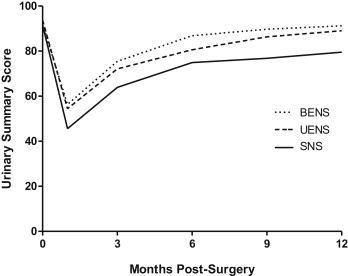
A total of 102 preoperatively potent men underwent laparoscopic or robotic radical prostatectomy; NS was prospectively graded at surgery using a 0-4 scale/neurovascular bundle. Urinary functional outcomes were measured by validated Expanded Prostate Cancer Index Composite questionnaire at baseline and follow-up time points (1, 3, 6, 9, and 12 months) in 99 men who underwent various degrees of NS. Mixed linear regression was used to analyze the effect of NS quality and other clinical factors on urinary outcomes.

RESULTS:

Patients with at least 1 neurovascular bundle spared completely, along with its supportive tissues (NS grade 4/4), noted significantly improved Expanded Prostate Cancer Index Composite urinary functional and continence outcomes as early as 1 month postoperatively and up to 12 months. Significantly less urinary bother was also noted in these men by 9-12 months postoperatively. Multivariate analysis revealed that bilateral or unilateral excellent NS (at least 1 bundle graded 4/4), increasing time from surgery, young patient age, and lower body mass index positively and significantly affected urinary functional outcomes, including pad use. Men who received excellent unilateral NS recovered urinary function about as well as men who had both neurovascular bundles spared in similar fashion.

CONCLUSION:

The quality of NS significantly influences patient-defined urinary functional convalescence. Completely sparing at least 1 neurovascular bundle along with its supportive tissues has a dramatic effect on the recovery of urinary continence and quality of life in preoperatively potent men.


PubMed

Download PDF


Physician in operating room

Prostate Cancer Risk Calculator for MRI-guided biopsy (PCRC-MRI)

Original Story Published in Folio Magazine
February 03, 2022 By Adrianna MacPherson

Risk calculator helps pinpoint which patients are suitable for MRI-guided prostate biopsies that improve on the current standard of care.

Adam Kinnaird, a surgeon and assistant professor in the Faculty of Medicine & Dentistry, and a member of both APCaRI and CRINA collaborated with colleagues at UCLA to develop a new risk calculator that could reduce the number of unnecessary and invasive biopsies for prostate cancer. Dr. Kinnaird explains that the tool is available online for free, requires only conventional clinical data paired with data from prostate MRI scans, and targets an unmet clinical need for prostate cancer patients.

The efficacy and accuracy of the calculator were calculated from a 10-year study Kinnaird led on 2,354 men undergoing MRI-guided prostate biopsy. The results, published in CUAJ on October 18th 2021,  showed that the simple, free calculator significantly helped improve patient selection for biopsies.

More than one million prostate biopsies are performed in North America every year, and prostate cancer is the most common type of internal cancer in North American men. However, as Kinnaird explained, these biopsies aren’t always necessary and they come with certain risks.

“Only about 25 to 30 percent of them may show some prostate cancer, and up to seven percent of men, after they’ve had a prostate biopsy, end up getting hospitalized within 30 days. And, there’s a four percent risk of what’s called post-biopsy sepsis, which can cause patients to end up in the ICU or may even prove fatal,” said Kinnaird. “Anything we can do before the biopsy to determine whether the patient really needs a biopsy is very important for patient care.”

The risk calculator, called the PCRC-MRI, is the first in North America developed to predict biopsy outcomes in North American men. The combination of the standard-of-care data with MRI data provides two critical benefits: it triages patients to determine whether they truly need the biopsy in the first place, and if they do, it improves the technique so clinicians are better able to find more clinically significant prostate cancer.

“We did a net benefit analysis using this calculator. The analysis showed that we could safely avoid 16 biopsies out of every 100 at a risk threshold of 20 percent. What that means is we could potentially be saving 16 men from having to undergo this invasive procedure that has serious potential side effects,” said Kinnaird.

Kinnaird noted that the MRI-guided prostate biopsy is already a major improvement on the current standard of care in Alberta, called a 12-core transrectal ultrasound-guided biopsy.

“It’s basically a blind biopsy. An ultrasound probe can see the prostate, it can see the outline, but it cannot see if there’s actually any prostate cancer in the prostate,” Kinnaird explained. “So what’s done is the urologist or radiologist performing the biopsy takes 12 random samples from the prostate and hopes that they detect prostate cancer if it’s there.”

While many of the patients in the study received MRI-guided prostate biopsies at the University of California, Los Angeles, the U of A recently obtained a device called the UroNAV that allows patients to receive the same type of biopsy and gives clinicians a new tool in their arsenal.

“It gives us something to aim at,” said Kinnaird. “We take what we’ve learned from the MRI being done on the patient and create a model of their prostate in the software. Then we can fuse it to the ultrasound being done in real-time. It’s been shown to improve the detection of clinically significant prostate cancer and to reduce the rate or risk of identifying clinically insignificant prostate cancer, which is prostate cancer that we would not want to treat and therefore not want to detect.”

For the clinical data component, the PCRC-MRI examines certain variables that have previously been identified as risk factors for harbouring prostate cancer, from family history to an abnormal digital rectal examination.

The next steps involve creating more calculators that provide clinicians with beneficial information that can help with selecting patients for these invasive biopsies. Kinnaird is now working on a risk calculator for patients under active surveillance, which would examine the likelihood of their low-risk prostate cancer becoming more aggressive.

The study was supported by funding from the U.S. National Cancer Institute and the UCLA Clinical and Translational Science Institute.

- Perrin Beatty