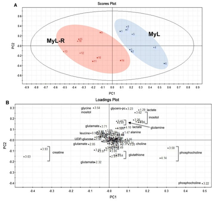
There has been a recent shift in how cancers are defined, where tumors are no longer simply classified by their tissue origin, but also by their molecular characteristics. Furthermore, personalized medicine has become a popular term and it could start to play an important role in future medical care. However, today, a “one size fits all” approach is still the most common form of cancertreatment. In this mini-review paper, we report on the role of nuclear magneticresonance (NMR) metabolomics in drug development and in personalized medicine. NMR spectroscopy has successfully been used to evaluate current and potential therapies, both single-agents and combination therapies, to analyze toxicology, optimal dose, resistance, sensitivity, and biological mechanisms. It can also provide biological insight on tumor subtypes and their different responses to drugs, and indicate which patients are most likely to experience off-target effects and predict characteristics for treatment efficacy. Identifying pre-treatmentmetabolic profiles that correlate to these events could significantly improve how we view and treat tumors. We also briefly discuss several targetedcancerdrugs that have been studied by metabolomics. We conclude that NMR technology provides a key platform in metabolomics that is well-positioned to play a crucial role in realizing the ultimate goal of better tailored cancer medicine.
Konstantin Stoletov and Lian Willetts co-first-authored an article published recently in Nature Communications titled “Quantitative in vivo whole genome motility screen reveals novel therapeutic targets to block cancer metastasis“. These two researchers, along with fellow Lewis lab members and collaborators from the University of Calgary and Vanderbilt University set out to determine what genes and signaling networks are involved in the rate-limiting steps of solid tumour cell motility, in vivo. But the team was hampered by the lack of an effective, quantitative, in vivo imaging platform. They wanted to visualize the movement of tumour cells, or lack of, in real-time AND use this intravital imaging platform to screen a large bank of tumour cells harboring single gene mutations for cells that show a loss of motility.
When tumour cells metastasize they get into (intravasate) the hosts’ bloodstream and use the vascular system like roadways to travel throughout the body. This lets the tumour cells colonize new microenvironments where they will proliferate and form new tumours. So metastatis is really dependent on tumour cell motility. Although there are many different types of solid tumours known, previous research suggests that if the tumour cells can mobilize and metastasize then the expressed motility-related genes share homology across tumour types. This is great news because it would mean that therapeutic targets aimed at stopping motility could also stop metastasis for many tumour types!
Dr. Konstantin Stoletov
Dr. Lian Willetts
The Lewis lab researchers and their collaborators developed an in vivo, fluorescent, time-lapse screening platform that uses shell-less avian embryos for tumour growth and formation. The avian embryo is an excellent tumour model because the tumour cells will grow on the chorioallantoic membrane in a single cell layer, making in vivo cell motility imaging actually doable.
Using this platform the team screened over 30 000 human genes for the ones needed for cell motility and ultimately found 17 genes that looked to be effective metastasis-blocking gene targets. Stoletov, along with other Lewis lab members, are continuing this research by studying these 17 attractive candidates further to determine which one (s) would make therapeutic metastasis-blocking targets.
This article has generated a lot of interest in the scientific community and in the general public! Check out the links below to mentions and articles in the media.