Publications

Publications

ERG protein expression and gene rearrangements are present at lower rates in metastatic and locally advanced castration-resistant prostate cancer compared to localized disease.

By:
Contributors: Bryan Donnelly, MD, MSc, FRCSC, Kiril Trpkov, MD, FRCPC, Tarek Bismar Research Group
Urology. 2013 Aug;82(2):394-9. doi: 10.1016/j.urology.2013.03.029. Epub 2013 Jun 6.

Abstract


OBJECTIVE:

To compare ERG expression and gene rearrangements rates in metastatic and castrationresistant prostate cancer (CRPC) to localized disease as ERG is the most common genetic event in early prostate cancer (PCa) with potential prognostic and therapeutic implications.

METHODS:

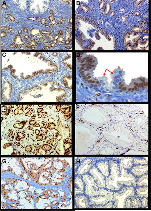
We evaluated ERG protein expression in 344 patients with PCa in 3 cohorts including localized, metastatic, and castrationresistant disease using immunohistochemistry (IHC) and fluorescence in situ hybridization (FISH).

RESULTS:

ERG protein expression was detected exclusively in the neoplastic epithelium and was found in 6.8% and 46.3% of high-grade prostatic intraepithelial neoplasia (HGPIN) and localized PCa, respectively. In metastatic and locally advanced CRPC, ERG expression was significantly lower, occurring at 36.1% and 37.2%, respectively. In PCa with foamy gland morphology, ERG protein expression was detected in only 18.6% compared with reported rates of about 42%-48% in acinar PCa. Moreover, ERG protein expression and gene rearrangements showed an overall consistency rate of 90.6% (P <.0001). The consistency rate was 100% both in benign glands and HGPIN, and 96.1% in localized PCa. However, it was significantly lower at 76.9% and 85% in node metastatic and CRPC, respectively (P <.0001).

CONCLUSION:

ERG protein expression is restricted to neoplastic prostatic epithelium and is present at lower rates in metastatic and CRPC compared to localized PCa. IHC and FISH concordance rates were significantly lower in node metastatic and CRPC compared to localized PCa, which may suggest different biological and therapeutic implications. The lower rate of ERG protein expression in foamy gland PCa may suggest potential differences for this pattern of PCa at the molecular level.


PubMed

Download PDF

Why did the chicken cross the road…?

Konstantin Stoletov and Lian Willetts co-first-authored an article published recently in Nature Communications titled “Quantitative in vivo whole genome motility screen reveals novel therapeutic targets to block cancer metastasis“. These two researchers, along with fellow Lewis lab members and collaborators from the University of Calgary and Vanderbilt University set out to determine what genes and signaling networks are involved in the rate-limiting steps of solid tumour cell motility, in vivo. But the team was hampered by the lack of an effective, quantitative, in vivo imaging platform. They wanted to visualize the movement of tumour cells, or lack of, in real-time AND use this intravital imaging platform to screen a large bank of tumour cells harboring single gene mutations for cells that show a loss of motility.

https://www.nature.com/articles/s41467-018-04743-2

When tumour cells metastasize they get into (intravasate) the hosts’ bloodstream and use the vascular system like roadways to travel throughout the body. This lets the tumour cells colonize new microenvironments where they will proliferate and form new tumours. So metastatis is really dependent on tumour cell motility. Although there are many different types of solid tumours known, previous research suggests that if the tumour cells can mobilize and metastasize then the expressed motility-related genes share homology across tumour types. This is great news because it would mean that therapeutic targets aimed at stopping motility could also stop metastasis for many tumour types!

Dr. Konstantin Stoletov


Dr. Lian Willetts
Dr. Lian Willetts

The Lewis lab researchers and their collaborators developed an in vivo, fluorescent, time-lapse screening platform that uses shell-less avian embryos for tumour growth and formation. The avian embryo is an excellent tumour model because the tumour cells will grow on the chorioallantoic membrane in a single cell layer, making in vivo cell motility imaging actually doable.

Using this platform the team screened over 30 000 human genes for the ones needed for cell motility and ultimately found 17 genes that looked to be effective metastasis-blocking gene targets. Stoletov, along with other Lewis lab members, are continuing this research by studying these 17 attractive candidates further to determine which one (s) would make therapeutic metastasis-blocking targets.

This article has generated a lot of interest in the scientific community and in the general public! Check out the links below to mentions and articles in the media.

https://www.biocentury.com/bc-innovations/translation-brief/2018-07-18/how-chicken-embryo-screen-identified-entos%E2%80%99-

https://www.ualberta.ca/medicine/news/2018/june/putting-the-brakes-on-metastatic-cancer

Stay tuned for a podcast that will be posted soon from “Parsing Science” where the hosts interview Dr. John Lewis about this work!

https://www.parsingscience.org/coming_soon/
UPDATE Oct 12, 2018: The podcast with John Lewis on Parsing Science called “Halting Cancers’ Spread“, is now available!

- Perrin Beatty