Publications

Publications

Comparing efficiency of micro-RNA and mRNA biomarker liberation with microbubble-enhanced ultrasound exposure.

By:
Contributors: Robert Paproski, PhD
Ultrasound Med Biol. 2014 Sep;40(9):2207-16. doi: 10.1016/j.ultrasmedbio.2014.05.005. Epub 2014 Jul 9.

Abstract

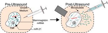
Blood biomarkers are potentially powerful diagnostic tools that are limited clinically by low concentrations, the inability to determine biomarker origin and unknown patient baseline. Recently, ultrasound has been shown to liberate proteins and large mRNA biomarkers, overcoming many of these limitations. We have since demonstrated that adding lipid-stabilized microbubbles elevates mRNA concentration an order of magnitude compared with ultrasound without microbubbles, in vitro. Unfortunately the large size of some mRNA molecules may limit efficiency of release and hinder efficacy as an ultrasound-liberated biomarker. We hypothesize that smaller molecules will be released more efficiently with ultrasound than larger molecules. Although investigation of large libraries of biomarkers should be performed to fully validate this hypothesis, we focus on a small subset of mRNA and micro-RNAs. Specifically, we focus on miR-21 (22 base pairs [bp]), which is upregulated in certain forms of cancer, compared with previously investigated mammaglobin mRNA (502 bp). We also report release of micro-RNA miR-155 (22 bp) and housekeeping rRNA S18 (1869 bp). More than 10 million additional miR-21 copies per 100,000 cells are released with ultrasound-microbubble exposure. The low- molecular-weight miR-21 proved to be liberated 50 times more efficiently than high-molecular-weight mammaglobin mRNA, releasing orders of magnitude more miR-21 than mammaglobin mRNA under comparable conditions.

 PubMed

Download PDF

The Calgary Prostate Cancer Centre has the highest accrual for a novel ultrasound study in prostate cancer

“We have enrolled over 400 patients at our site, reaching our enrollment goal much faster than all other sites across North America. We are now planning on adding in 250 more patients to this trial because of the encouraging results found with the first arm of the trial. Our site tied with the highest accrual goal and surpassed all other sites to meet our enrollment goal.”

The study is a “Multi-Center trial of high-resolution transrectal ultrasound versus standard low-resolution transrectal ultrasound for the identification of clinically significant prostate cancer”

The only definitive method for diagnosing prostate cancer is through a prostate biopsy. This procedure includes the use of an ultrasound machine to guide both freezing needles and biopsy needles into the prostate. The ultrasound machine that is currently in use is a low-resolution ultrasound machine which means that although it is good at seeing the entire prostate gland to guide the needles, it is often unable to visualize the prostate in enough detail to be able to see different lesions and areas of concern within it. Thus, many biopsy samples are taken systematically with two samples from each section of the prostate. Recently a new ultrasound machine has been created that gives images of the prostate with much higher resolution, allowing the radiologist performing the biopsy to see details within the prostate that were previously inaccessible. A study using this new high-resolution ultrasound machine is being completed at the Prostate Cancer Centre to compare the adequacy of this new machine to detect prostate cancer over the standard low-resolution machine. Over 650 patients will be enrolled in this study!

pcc-logo_2x

- Eric Hyndman