Publications

Publications

Enhanced Detection of Cancer Biomarkers in Blood-Borne Extracellular Vesicles Using Nanodroplets and Focused Ultrasound

Cancer Res. 2017 Jan 1;77(1):3-1

Paproski RJ, Jovel J, Wong GK, Lewis JD, Zemp RJ.

Abstract

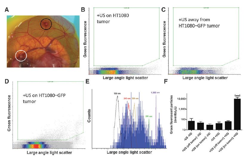
The feasibility of personalized medicine approaches will be greatly improved by the development of noninvasive methods to interrogate tumor biology. Extracellular vesicles shed by solid tumors into the bloodstream have been under recent investigation as a source of tumor-derived biomarkers such as proteins and nucleic acids. We report here an approach using submicrometer perfluorobutane nanodroplets and focused ultrasound to enhance the release of extracellular vesicles from specific locations in tumors into the blood. The released extracellular vesicles were enumerated and characterized using micro flow cytometry. Only in the presence of nanodroplets could ultrasound release appreciable levels of tumor-derived vesicles into the blood. Sonication of HT1080-GFP tumors did not increase the number of circulating tumor cells or the metastatic burden in the tumor-bearing embryos. A variety of biological molecules were successfully detected in tumor-derived extracellular vesicles, including cancer-associated proteins, mRNAs, and miRNAs. Sonication of xenograft HT1080 fibrosarcoma tumors released extracellular vesicles that contained detectable RAC1 mRNA with the highly tumorigenic N92I mutation known to exist in HT1080 cells. Deep sequencing serum samples of embryos with sonicated tumors allowed the identification of an additional 13 known heterozygous mutations in HT1080 cells. Applying ultrasound to HT1080 tumors increased tumor-derived DNA in the serum by two orders of magnitude. This work is the first demonstration of enhanced extracellular vesicle release by ultrasound stimulation and suggests that nanodroplets/ultrasound offers promise for genetic profiling of tumor phenotype and aggressiveness by stimulating the release of extracellular vesicles.

PubMed

Download PDF

Why did the chicken cross the road…?

Konstantin Stoletov and Lian Willetts co-first-authored an article published recently in Nature Communications titled “Quantitative in vivo whole genome motility screen reveals novel therapeutic targets to block cancer metastasis“. These two researchers, along with fellow Lewis lab members and collaborators from the University of Calgary and Vanderbilt University set out to determine what genes and signaling networks are involved in the rate-limiting steps of solid tumour cell motility, in vivo. But the team was hampered by the lack of an effective, quantitative, in vivo imaging platform. They wanted to visualize the movement of tumour cells, or lack of, in real-time AND use this intravital imaging platform to screen a large bank of tumour cells harboring single gene mutations for cells that show a loss of motility.

https://www.nature.com/articles/s41467-018-04743-2

When tumour cells metastasize they get into (intravasate) the hosts’ bloodstream and use the vascular system like roadways to travel throughout the body. This lets the tumour cells colonize new microenvironments where they will proliferate and form new tumours. So metastatis is really dependent on tumour cell motility. Although there are many different types of solid tumours known, previous research suggests that if the tumour cells can mobilize and metastasize then the expressed motility-related genes share homology across tumour types. This is great news because it would mean that therapeutic targets aimed at stopping motility could also stop metastasis for many tumour types!

Dr. Konstantin Stoletov


Dr. Lian Willetts
Dr. Lian Willetts

The Lewis lab researchers and their collaborators developed an in vivo, fluorescent, time-lapse screening platform that uses shell-less avian embryos for tumour growth and formation. The avian embryo is an excellent tumour model because the tumour cells will grow on the chorioallantoic membrane in a single cell layer, making in vivo cell motility imaging actually doable.

Using this platform the team screened over 30 000 human genes for the ones needed for cell motility and ultimately found 17 genes that looked to be effective metastasis-blocking gene targets. Stoletov, along with other Lewis lab members, are continuing this research by studying these 17 attractive candidates further to determine which one (s) would make therapeutic metastasis-blocking targets.

This article has generated a lot of interest in the scientific community and in the general public! Check out the links below to mentions and articles in the media.

https://www.biocentury.com/bc-innovations/translation-brief/2018-07-18/how-chicken-embryo-screen-identified-entos%E2%80%99-

https://www.ualberta.ca/medicine/news/2018/june/putting-the-brakes-on-metastatic-cancer

Stay tuned for a podcast that will be posted soon from “Parsing Science” where the hosts interview Dr. John Lewis about this work!

https://www.parsingscience.org/coming_soon/
UPDATE Oct 12, 2018: The podcast with John Lewis on Parsing Science called “Halting Cancers’ Spread“, is now available!

- Perrin Beatty